Anterior Tibial Tuberosity Transfer Patella Realignment

What is the problem?

The patella is unstable and repeatedly dislocates. This is known as recurrent patellar dislocation. This pathology can be exacerbated by the abnormal position of the patellar tendon insertion on the anterior tibial tuberosity (ATT). This tendon connects the patella to the tibia where the insertion may be too lateral and/or too high resulting in the patella being in a position that increases the risk of dislocation.

Patella slightly tilted and off-centre

When should an anterior tibial tuberosity transfer be considered?

In the case of recurrent patella dislocations exacerbated by an abnormally lateral and/or higher position of the anterior tibial tuberosity, the ATT can be repositioned to stabilise the patella.

Lateral patellar dislocation

Before the operation

An ATT requires an operation planned during pre-surgery consultations based on knee x-rays and a CT scan.

Before the operation, a pre-anaesthesia consultation is conducted to check the patient is physically apt to undergo the operation and minimise the risk of postoperative complications.

The operation

The operation takes place in an operating theatre in compliance with strict standards of cleanliness and safety. The patient is placed supine on an operating table and a tourniquet is placed around the thigh. The operation lasts about 45 minutes and can be carried out under general or spinal anaesthesia. The latter is a regional anaesthetic anaesthetising the lower part of the body (as with an epidural). The anaesthetist will decide on the most suitable anaesthetic together with the patient.

A knee arthroscopy can be carried out before the ATT transfer to check the condition of the joint cartilage and remove any cartilage fragments fractured during the patellar dislocations. The lateral patellar retinaculum, the lateral attachment of the patella that tends to pull it outwards thus contributing to dislocation, is also cut.

The ATT is cut, repositioned correctly, and attached with 2 screws.

Final appearance: front view
Final appearance: side view
Final appearance: upper view

At the end of the procedure, the stability of the patella is tested and it must not dislocate.

No patella dislocation when bending

After the operation

A drain is inserted in the incision for several days to prevent the formation of a haematoma.

After the operation, the knee is partially numbed with a nerve block to help improve postoperative pain management. The effect lasts approximately 18 hours after the operation but can be extended with the insertion of a catheter in the thigh if the anaesthetist considers it necessary. Medication and ice on the knee also provide effective postoperative pain management.
After the operation, the knee is immobilised in a brace that must be worn for 6 weeks to allow the ATT to heal. Weight-bearing on the leg is permitted with the brace, possibly aided with crutches.

The stay in hospital is often between 1 and 3 days.

In general, two follow-up consultations will be necessary to monitor the healing of the osteotomy and recovery of the knee.

The duration of medical leave depends on the patient’s occupation and is often between 6 and 8 weeks.

Driving is possible again after 6 weeks. Sport can generally be resumed after 4 months.

Ultimately, the heads of the two screws used to attach the ATT may be uncomfortable when kneeling as they are just under the skin. It is not unusual to have them removed. This requires a short outpatient procedure (hospital stay of 1 day) and is usually performed after at least one year.

Risks linked to the operation

Unfortunately, zero risk does not exist in surgery. Any operation has its risks and limitations, which you must accept or not undergo the operation. However, if an operation is proposed, the surgeon and the anaesthetist consider that the expected benefits far outweigh the risk incurred.

Some risks, such as microbial infections of the surgical site, are common to all types of surgery. This complication is fortunately rare but when it occurs requires another operation and a course of antibiotics.

Bruising can also appear around the surgical site. This is usually prevented or reduced with a suction drain inserted at the end of the operation and removed in the days following the operation.

Knee surgery and lack of weight-bearing on the limb operated on also increase the risk of phlebitis, which can lead to a pulmonary embolism. To minimise this risk, blood thinners (in the form of daily injections or tablets) are prescribed for 3 weeks following the operation.

Failure of the osteotomy to fuse is called nonunion and requires further surgery to achieve healing of the ATT.

In rare cases, the knee remains stiff, hot, and painful for several months after the operation. This complication, known as algodystrophy or Chronic Regional Pain Syndrome (CRPS), is unpredictable and sometimes takes a long time to heal.

Finally, rarer complications can also occur. Nerves can also be damaged accidentally during the operation with a risk of paralysis or loss of feeling in the limb operated on, which can be transitory or permanent.

If you have any concerns about the operation, do not hesitate to talk to your surgeon or the anaesthetist and they will answer any questions you may have.

In summary...

When is surgery necessasry?

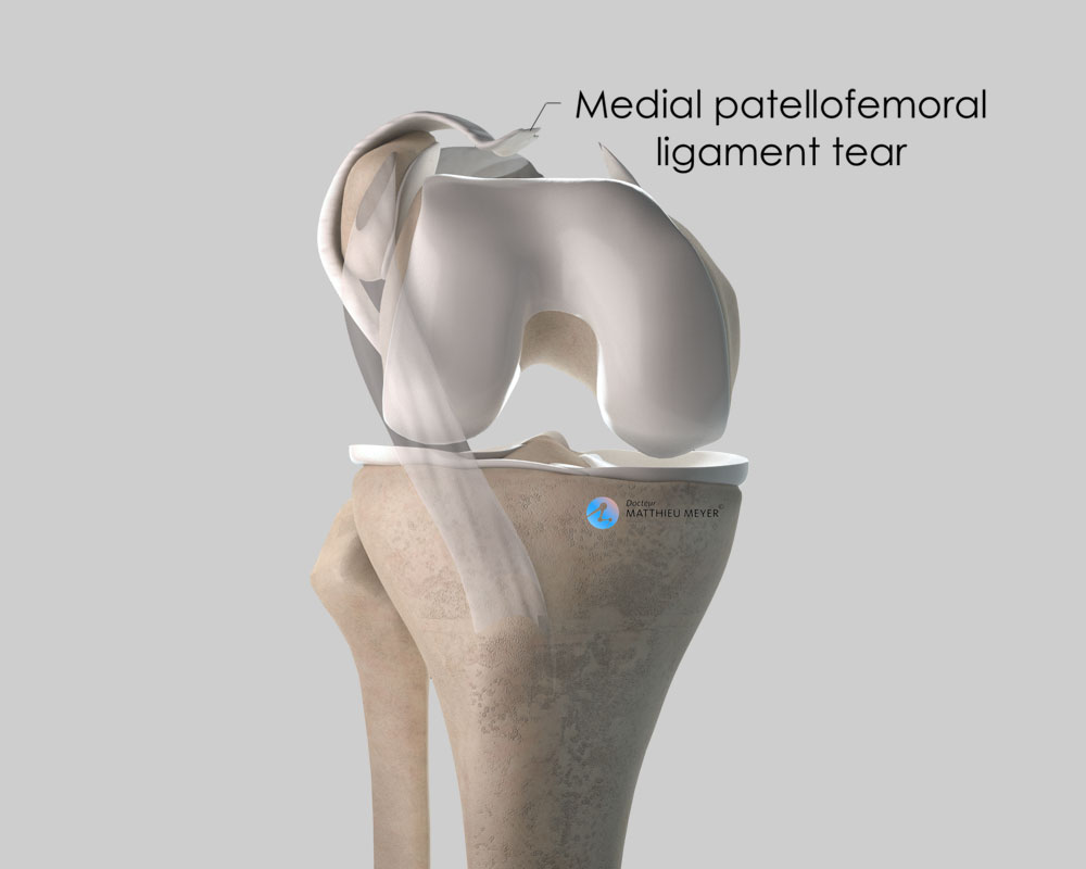
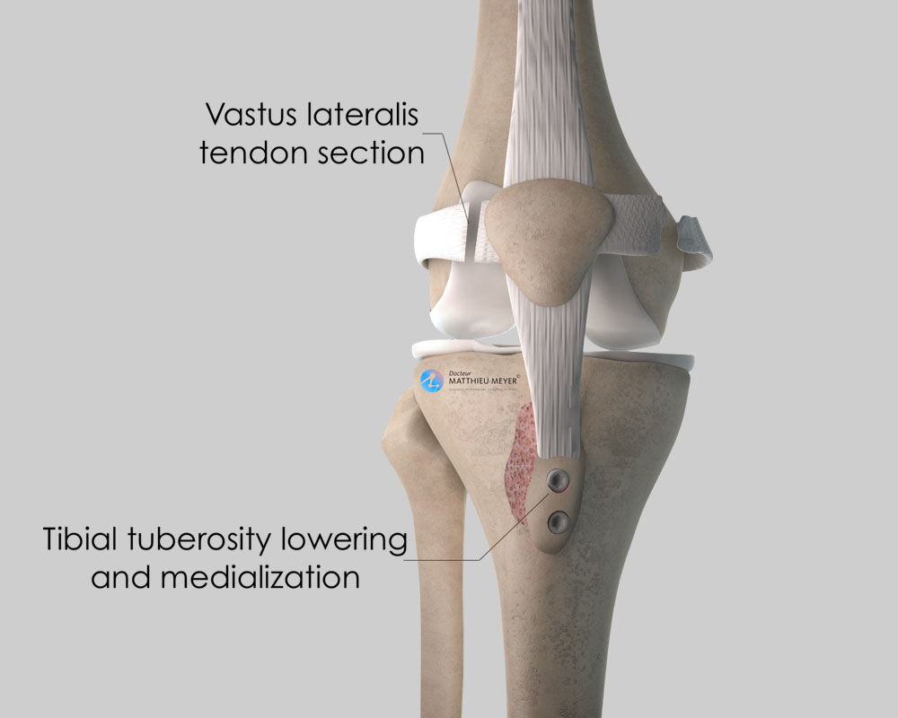
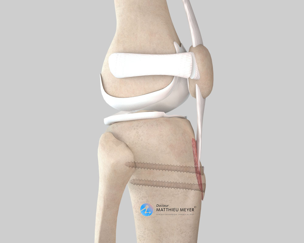
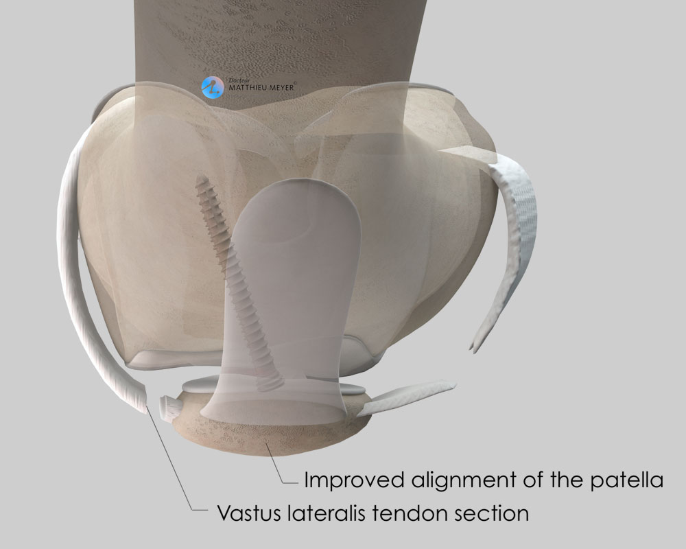
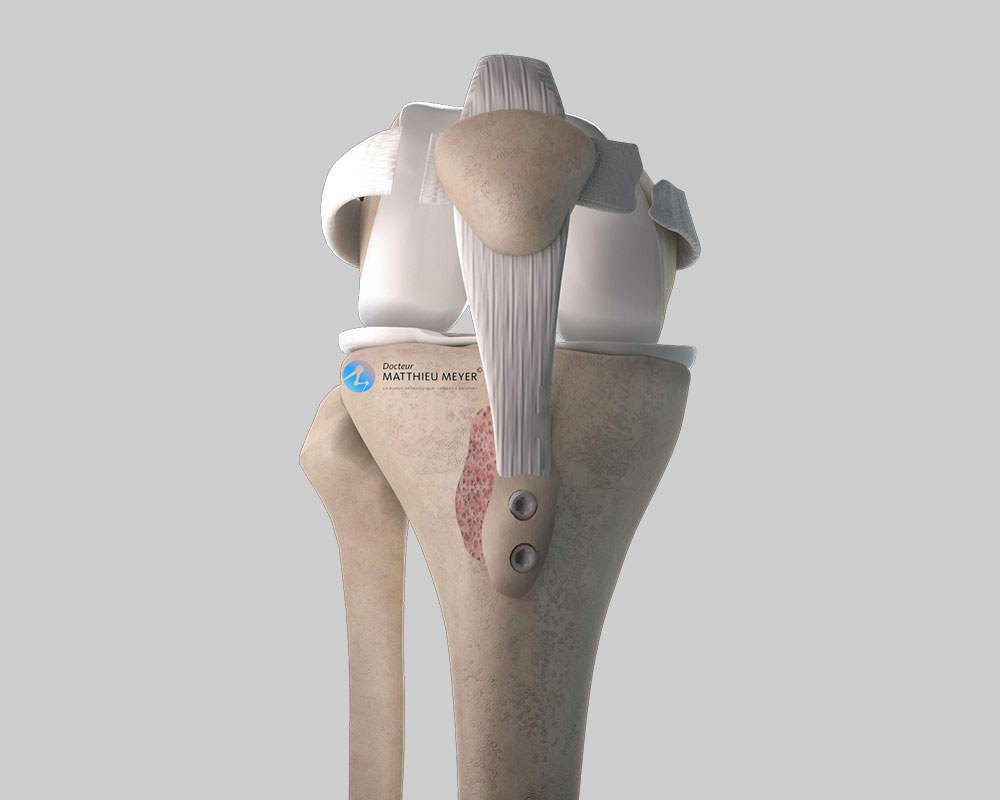
In the case of recurrent patellar dislocations, when the position of the ATT is too lateral and/or too high

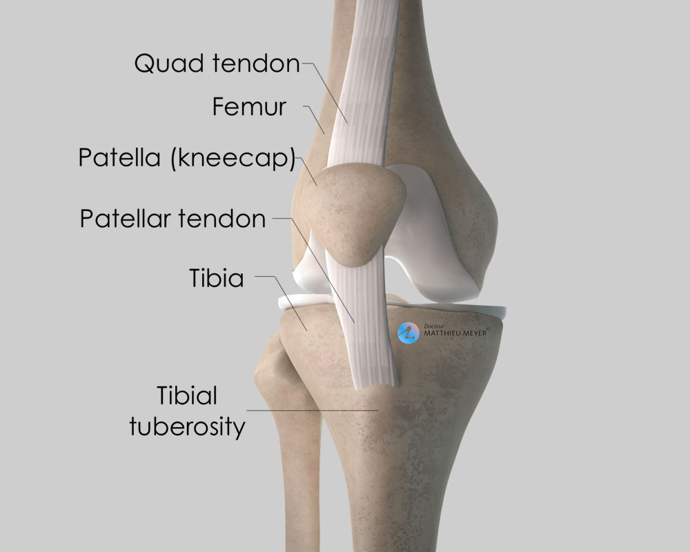
Aim of the operation

Stabilise the patella

Which anaesthesia?

General or regional (determined with the anaesthetist)

Duration of hospitalisation

Between 1 and 3 days

Resumption of weight-bearing

Immediately with a brace immobilising the knee

After the operation

Return home

Duration of rehabilitation

Generally 2 to 3 months

Duration of medical leave

6 to 8 weeks, longer for heavy work

Resumption of car driving

1½ months after the operation

Resumption of sport

4 months after the operation

Make an appointment

If you have any questions or wish to book a consultation,
please do not hesitate to contact us or make an appointment online via DoctoLib
Scroll to Top